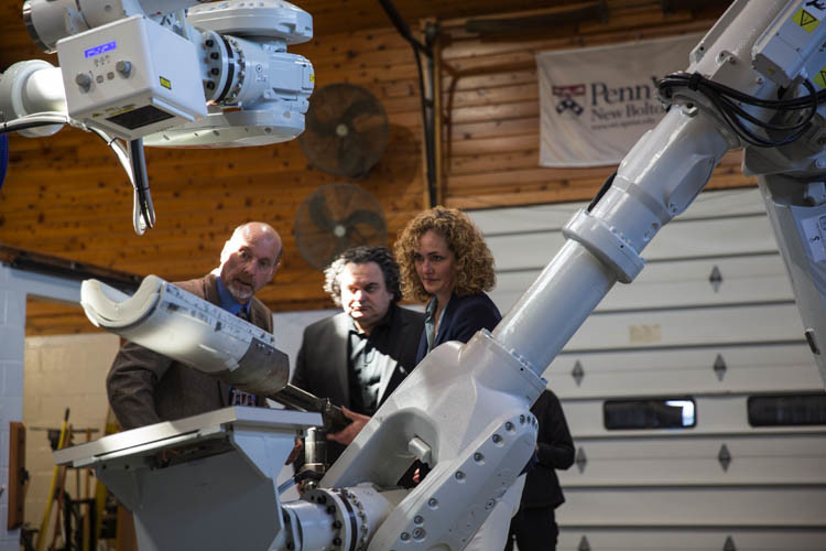
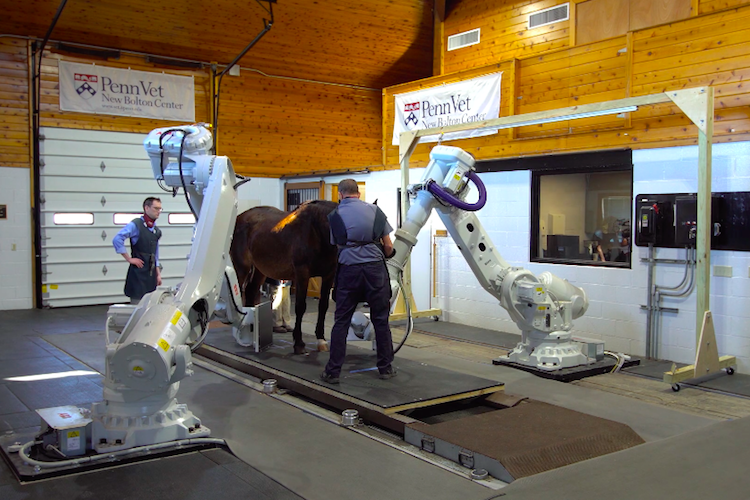
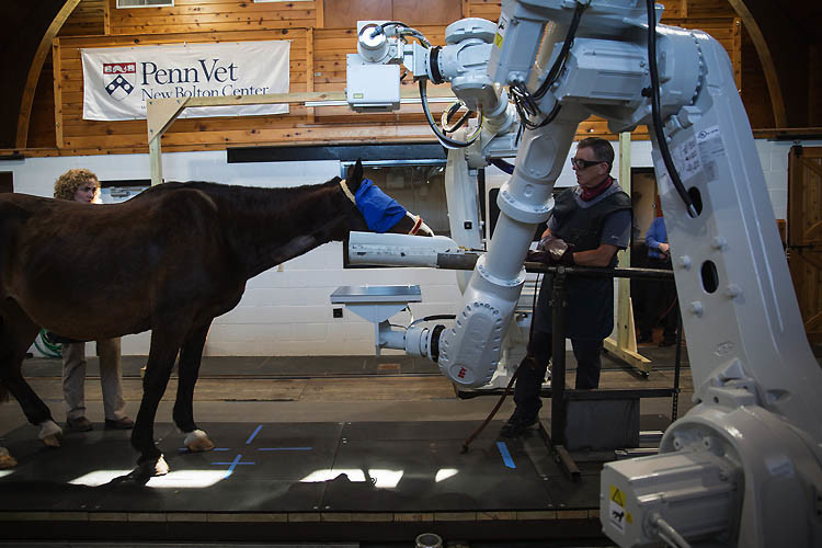
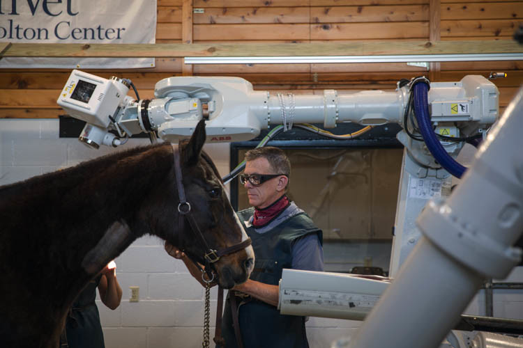
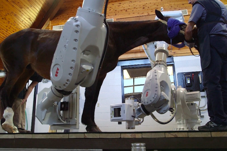
To make this possible, Penn Vet partnered with an imaging technology company called 4DDI to create the so called “Equimagine” scanner. The system uses a series of robots that can move around a standing patient to capture a variety of high-resolution images. In addition to the two-dimensional images popular in CT scanning, Equimagine can also capture fluoroscopic images (think short x-ray movies), three-dimensional scans, and radiographs at speeds of up to 16,000 frames per second.
Thoroughbred racehorses in particular commonly develop stress fractures that can be difficult to detect. When hairline fractures intensify enough for experts to notice the pain in the movements of the horse, it’s often too late to reverse the damage. “This technology has the potential to help diagnose those early enough that we can manage them and help prevent the horse from suffering a catastrophic breakdown on the race track,” said Dean Richardson, chief of large animal surgery at the New Bolton Center at Penn Vet.
Experts believe that adapting the horse-scanning technology for use in childhood medical imaging could even revolutionize pediatrics. Imagine a small child in need of a CT scan, hanging out while talking to his parents instead of terrified and on his back in a dark and lonely imaging tube.
In the future, veterinary researchers hope to be able to use the Equimagine system to capture motion images of a horse running on a treadmill, for example. The system’s programmable robots could enable a kind of medical imaging that has never been possible before, either in horses or in humans. “From a clinical standpoint, we will see elements of the horse’s anatomy that we’ve never seen before,” said Barbara Dallap Schaer, medical director of the New Bolton Center.
