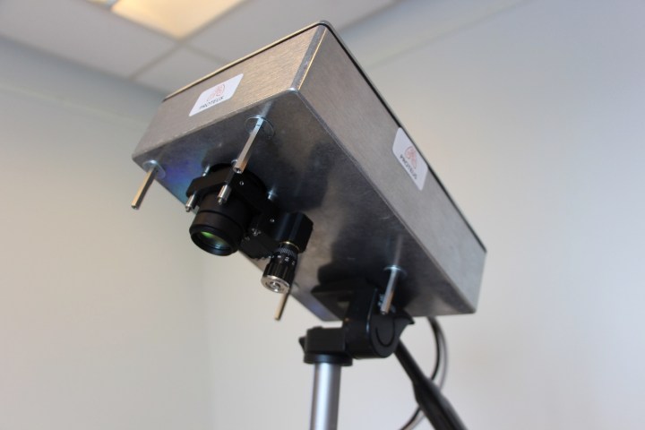
New medical imaging camera in the U.K. can see through the human body

medical camera human body spots illumination
Scientists at the U.K.’s University of Edinburgh recently developed an imaging medical camera that is capable of seeing through the human body to detect light sources. The camera is designed to help doctors better track medical tools called endoscopes, which are used to investigate a range of internal conditions. It can do this by detecting the illuminated end of the flexible endoscope cable.

“We have developed a method to determine the location of medical devices through tissue using an advanced camera technology,” Dr. Mike Tanner, a research fellow at Edinburgh’s Queens Medical Research Institute, told Digital Trends. “The principle is that some light does pass through tissue, as seen when holding a torch behind your hand. However, the amount of light passing through is very low, and the light scatters or bounces around inside the tissue structures losing all useful information. To solve this, we use a camera that is so sensitive it can see individual particles of light and also time the arrival of the photons. The very first light to arrive at the camera has been scattered least and tells us the location of the device.”

Up until now, physicians wanting to track the internal location of endoscopes have had to utilize expensive and potentially harmful scans such as X-rays to track the progress of the probes inside patients. The new imaging camera is able to change that, using thousands of single photon detectors packed onto a single silicon chip to track the location of light points through 20 centimeters of tissue under regular light conditions.

It is part of a wider initiative, in collaboration with Heriot-Watt University and the University of Bath, dedicated to developing new technologies for the diagnosis and treatment of lung disease.

“This [camera has already] been demonstrated in relevant scenarios, and we hope to take this forward to human trials in the coming year,” Tanner said. “The equipment is relatively simple and compact, and ideal for deployment and commercialization in realistic timescales.”

A paper describing the work, “Ballistic and snake photon imaging for locating optical endomicroscopy fibers,” was published in the journal Biomedical Optics Express.

Editors' Recommendations

Luke Dormehl
I'm a UK-based tech writer covering Cool Tech at Digital Trends. I've also written for Fast Company, Wired, the Guardian…
New Nikon Coolpix long-zoom cameras can shoot 4K video
nikon coolpix long zoom b700 b500 a900 featured

In addition to its new premium DL-series, Nikon is introducing three new long-zoom (megazoom and super-zoom) cameras to its Coolpix lineup. Besides having a long optical zoom lens, the B700 ($500), B500 ($300), and A900 ($400) have Nikon’s new SnapBridge “always on” wireless connectivity, while some models can shoot 4K video. The cameras will go on sale in the spring.

With a traditional DSLR-like super-zoom body, the B700 uses a 1/2.3-inch 20.2-megapixel back-illuminated (BSI) CMOS sensor. It’s not a large sensor like the 1-inch variant used in the DL-series, but it’s typical of point-and-shoots. The lens has a 60x zoom with Dual Detect Vibration Reduction and 5-Axis Hybrid Vibration Reduction (optical and digital) for videos, and the camera can shoot 5 frames per second (fps) continuously. The B700 can shoot RAW photos and 4K videos at 30p. The 3-inch LCD is a vari-angle type that flips out and tilts into a variety of positions. ISO sensitivity ranges between 100-3,200. For more advanced users, there are several shooting modes and manual settings.

Read more
This tiny new radio module can wirelessly broadcast data from inside the human body
smart medical devices injectable radio

Implantable medical devices like pacemakers excel at regulating body processes, but they usually do so in a "dumb" manner without any computer analysis or communications support. While most researchers are looking to develop flexible and lifelike materials for medical devices, University of Michigan Professor David Blaauw is taking a different approach. He wants to improve the intelligence of these devices by equipping them with a miniature computing and communications system, reports IEEE Spectrum.

According to Blaauw, implantable electronics often lack the processing power to analyze the body in-situ, and do not include the communications hardware necessary to wirelessly send those results outside of the human body. The stumbling block is not the processor technology, which can be miniaturized fairly easily. Instead, the most difficult component to shrink is the radio module, which often is rather large due to its power and antenna requirements.

Read more
MIT researchers develop RF technology to see through walls, distinguish between humans
mit researchers want to use rf x ray vision improve motion capture save lives

A group of MIT researchers have built upon previous advances in the use of wireless signals to track human motion through walls to show how subtle human movements can be observed through objects to distinguish between individuals. There are implications for heating bills, motion capturing for movies, and keeping family members safe in a smart home.

A team of researchers (two professors and three students) at MIT’s Computer Science and Artificial Intelligence Lab (CSAIL) have constructed a device that uses radio frequency (RF) signals to take snapshots of a human figure through a wall, which are then pieced together in a “reconstruction algorithm” to generate a heat-map-like silhouette.

Read more